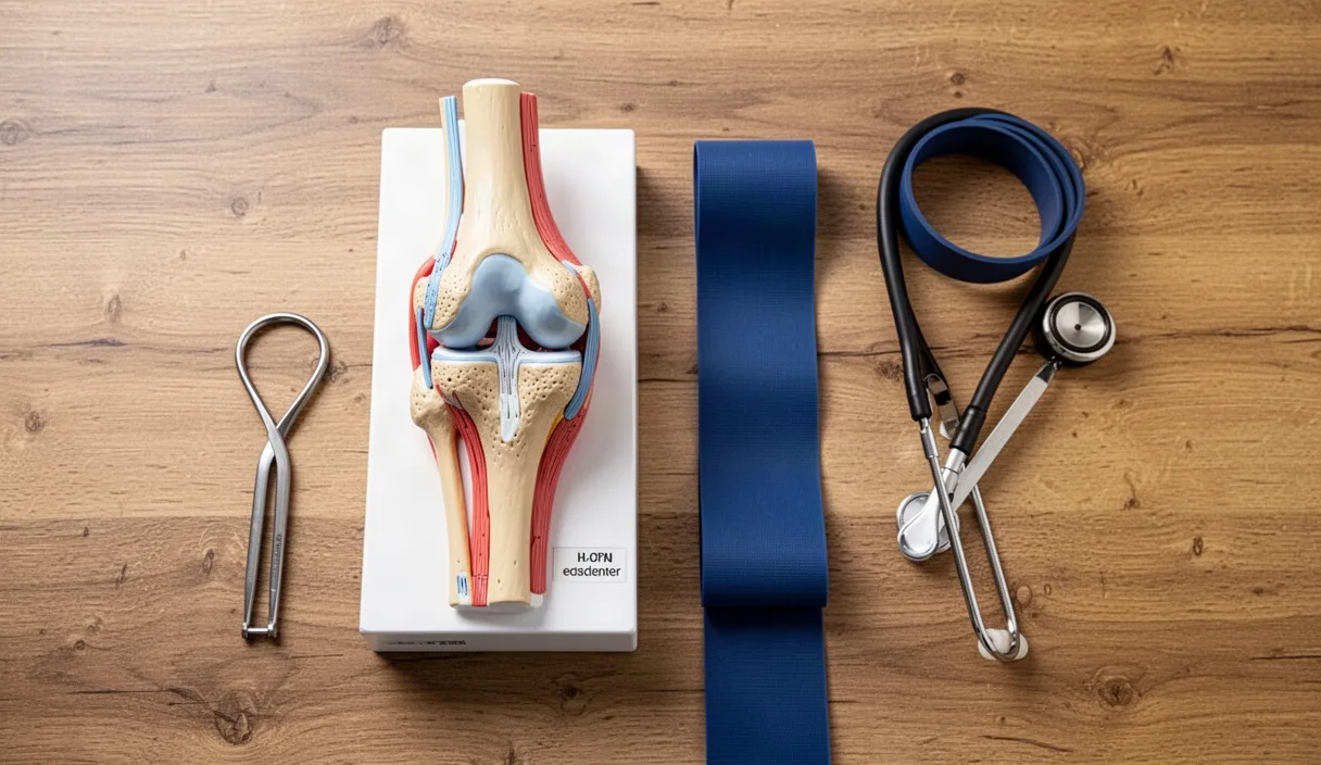
Cartilage covers the ends of bones where they meet to form a joint. In a healthy knee or hip, this tissue is white, smooth, and rubbery. It is between two and four millimeters thick in human knees. Its job is to absorb compressive forces and let bones glide past each other without friction. When cartilage wears thin or tears, bone rubs against bone, causing pain and restricted range of motion.
Many people believe that resting an aching joint protects the tissue. Long periods of bed rest or inactivity actually starve the tissue. Cartilage needs mechanical pressure to stay healthy. Mechanical load drives fluid into and out of the joint structure. Without this process, the cells that maintain the tissue die off, and the joint surface becomes brittle.
The avascular nature of articular cartilage
Most tissues in the human body heal through blood supply. Muscles, skin, and bones have deep networks of capillaries. When a muscle tears, blood delivers oxygen, amino acids, and white blood cells directly to the damage site. Articular cartilage does not have blood vessels. Medical professionals call this an avascular tissue. It also lacks nerves and a lymphatic system.
Chondrocytes are the only cells found in healthy cartilage. They produce and maintain the extracellular matrix, which consists of water, type II collagen, and proteoglycans. Because chondrocytes have no direct blood supply, they cannot receive glucose or oxygen from red blood cells. They survive in a low-oxygen environment. Their metabolic rate is slow compared to muscle tissue, which explains why cartilage takes months or years to respond to treatment instead of weeks.
Nutrients must reach chondrocytes from outside the tissue. The primary source of nutrients is synovial fluid, a viscous liquid that fills the joint capsule. Nutrients dissolved in this fluid must travel through the matrix by passive diffusion and convection. If the fluid remains static, nutrient delivery drops. The matrix begins to dry out, and the chondrocytes lose their ability to manufacture fresh collagen fibers.
Cartilage is divided into four distinct microscopic zones, each with a different structural role:
- Superficial zone: The top ten to twenty percent of the tissue. Collagen fibers lie flat, parallel to the joint surface, to resist shear forces from sliding bones.
- Middle zone: Thick collagen fibers run in diagonal patterns. This layer contains high levels of proteoglycans to absorb compression.
- Deep zone: Fibers run vertically, perpendicular to the bone. This zone provides the greatest resistance to direct downward loads.
- Calcified zone: Mineralized tissue that anchors the cartilage matrix directly to the subchondral bone beneath it.
The sponge mechanism of synovial fluid exchange
Articular cartilage works like a dense household sponge submerged in a bucket of water. When you squeeze the sponge, dirty water flows out. When you release the sponge, clean water flows back into the pores. Cartilage performs this exact exchange under intermittent mechanical pressure.
When you put body weight onto a joint, compressive pressure rises inside the cartilage matrix. Water and small waste products are forced out into the joint cavity. When you lift the foot or remove the weight, the pressure drops instantly. The proteoglycan molecules inside the matrix carry a negative electrical charge. These charges attract water molecules back into the matrix, pulling in dissolved glucose, oxygen, and amino acids from the synovial fluid.
This process is called fluid exudation and imbibition. It only works if the loading cycle alternates regularly between pressure and release. Constant compression keeps the fluid pressed out, leaving the cells without nutrients. Constant bed rest leaves the fluid sitting still, with no mechanical gradient to force nutrient exchange.
The rate of exchange depends on three physical factors:
- Deformation depth: The cartilage must compress by roughly ten to twenty percent of its resting height to drive fluid flow.
- Cycle frequency: Loading cycles between 0.5 and 1.5 hertz (about one step per second) produce the best balance of fluid exchange and tissue safety.
- Fluid viscosity: Synovial fluid thins out as joint temperature rises during movement, making it easier for nutrients to pass into the matrix.
Impact loading versus smooth cyclical movement
Not all physical loads affect joint tissue in the same way. Cartilage responds well to predictable, cyclical forces. It breaks down when exposed to sudden peak forces that exceed the tensile strength of the collagen framework.
Smooth cyclical movement includes walking on flat ground, riding a stationary bicycle, using a rowing machine, and water aerobics. In these activities, the joint moves through a defined range of motion. The compressive force builds gradually and releases predictably. This allows the fluid to flow without tearing the superficial collagen fibers.
Impact loading involves sudden changes in speed, jumping, hard landings, and rapid direction changes. When a foot strikes concrete during a sprint, the impact force can reach three to five times body weight in a fraction of a second. If the surrounding muscles are tired, the cartilage takes the brunt of that spike. A sudden spike can cause micro-cracks in the deep zone or shear the superficial layer away from the bone.
| Movement Type | Peak Force (x Body Weight) | Deformation Rate | Cartilage Response |
|---|---|---|---|
| Stationary cycling (moderate cadence) | 1.2 to 1.5 | Slow and regular | High fluid exchange, zero shear stress |
| Level ground walking (3 mph) | 2.1 to 2.8 | Moderate and regular | Good nutrient delivery, healthy matrix maintenance |
| Downhill hiking | 3.8 to 4.5 | Fast with high shear | Increased risk of superficial layer micro-trauma |
| Box jumps or court sports | 4.5 to 7.0 | Sudden peak spike | Potential matrix micro-fractures if unconditioned |
For an individual with cartilage wear, the goal is to choose movements with low peak forces and high repetition counts. This delivers the maximum amount of synovial fluid exchange without causing mechanical damage to the collagen matrix.
Managing load volume when joints feel stiff
Joint stiffness in the morning is a common complaint. It usually happens because synovial fluid thickens overnight while the joint sits cold and still. The urge is to sit down until the joint feels better. That approach prolongs the stiffness. You must apply low-intensity movement to warm the fluid and restart fluid circulation.
Use a graded exposure rule when scheduling your activity. If a joint aches, assess the pain on a scale from zero to ten. A mild discomfort between two and four is acceptable during movement, provided it settles back to your baseline within two hours after finishing. If your pain spikes above five, or if the joint swells that evening, you exceeded the capacity of the tissue. Reduce your volume on the next session.
To reduce joint stiffness before a walk or daily chores, follow these steps:
- Unloaded joint circles: Sit on a high chair with your feet off the floor. Swing your lower legs gently forward and back for two minutes. This stirs the synovial fluid without putting any body weight on the knee cartilage.
- Pelvic rocks: Lie on your back on a firm bed or floor mat. Draw your knees toward your chest until you feel mild pressure, then push them away. Repeat twenty times to lubricate the hip sockets.
- Short-stride walking: Begin your daily walk with shorter strides for the first four hundred meters. Short strides reduce the impact angle of the heel strike, lowering the force sent up through the ankle, knee, and hip.
- Cadence check: Aim for a step cadence of ninety to one hundred steps per minute. Shorter, quicker steps distribute the ground load across more cycles rather than fewer heavy strikes.
If a joint is hot to the touch, visibly swollen, or red, stop loaded exercise and consult a medical practitioner. Those symptoms indicate acute inflammation or infection, not standard mechanical stiffness. Loading an acutely inflamed joint can speed up enzymatic cartilage breakdown.
Recommended daily movement patterns for older adults
Older adults need consistent joint loading every day. Bone and cartilage turnover slows down with age, so long gaps between movement sessions lead to rapid tissue stiffening. A daily schedule should combine non-weight-bearing joint motion with controlled, low-impact loading.
Spend twenty to thirty minutes per day on continuous, cyclical loading. Walking on a flat path or using a pedal exerciser are two accessible options. Split this time into two shorter sessions if standing for twenty continuous minutes causes discomfort. Two ten-minute walks provide the same synovial fluid circulation as one twenty-minute walk, but with less cumulative joint fatigue.
Incorporate the following three movement patterns into your daily routine to maintain lower-body cartilage health:
- Sit-to-stand transitions: Sit on a firm, armless chair with your feet flat on the floor, shoulder-width apart. Lean your torso forward slightly and stand up without using your hands if possible. Lower yourself back down under control. Perform two sets of eight repetitions. This loads the hip and patellofemoral cartilage across their natural functional curves.
- Supported heel raises: Stand facing a kitchen counter. Place your fingertips on the counter for balance. Raise your heels off the floor over two seconds, hold for one second at the top, then lower over two seconds. Perform three sets of ten repetitions. This action compresses the ankle cartilage and strengthens the calf muscles that absorb walking shocks.
- Seated knee extensions: Sit tall against the back of a firm chair. Slowly straighten one knee until your leg is parallel to the floor. Hold your foot flexed for three seconds, then lower it over three seconds. Perform two sets of twelve repetitions per leg. This movement engages the quadriceps muscle and compresses the patellofemoral cartilage without body-weight impact.
Track your movement volume using a simple step counter or a kitchen timer. Consistency across seven days per week is far more valuable for cartilage health than one long, exhausting walk on the weekend.
Common mistakes
Many people make predictable errors when trying to manage joint health through movement. Recognizing these mistakes helps prevent avoidable joint irritation.
- Total rest during flare-ups: Sitting in a recliner for several consecutive days starves the tissue of synovial nutrients. Unless instructed by a physician, use unloaded movements like ankle pumps and seated leg swings instead of absolute stillness.
- Walking on hard, uneven surfaces: Concrete sidewalks and cobblestones deliver inconsistent impact angles to worn cartilage. Choose packed dirt paths, rubberized school tracks, or level asphalt.
- Ignoring footwear wear: Running and walking shoes lose their midsole shock absorption after roughly four hundred miles of use, even if the upper fabric looks new. Replace worn shoes to protect your joints from shock spikes.
- Sudden spikes in distance: Adding two miles to a walking routine overnight overloads cartilage before it can adapt. Increase your weekly walking time by no more than ten percent per week.
- Relying entirely on passive treatments: Heating pads, ice packs, and massage creams can alter pain signals, but they do not compress the matrix or move synovial fluid. Active, loaded movement is required for tissue nutrition.
Practical next steps
Start your cartilage health routine today with small, repeatable actions. Do not attempt an aggressive overhaul of your physical activity all at once. Cartilage requires weeks to adapt to structural changes in load.
First, test your joint response with a five-minute walk on a flat surface twice today. Note how your joints feel two hours later. If you experience no swelling or sharp pain, add two minutes to each walk tomorrow. Work your way up toward two fifteen-minute walks daily.
Second, clear a space near a sturdy chair or kitchen counter. Add the sit-to-stand and heel-raise exercises to your morning routine, right after breakfast. Make these movements a permanent daily habit, just like brushing your teeth.
Third, keep a simple written log on your refrigerator or nightstand. Record the minutes you walked and any joint symptoms you felt that evening. This log provides concrete data. If you consult a physical therapist, orthopedic specialist, or general practitioner, hand them this record. It will help them tailor a safe, long-term loading plan suited to your specific joint anatomy.